
Owl ears are uniquely built to help these birds hunt with remarkable accuracy, especially in the dark. Instead of visible outer ears, owls rely on hidden ear openings, a flexible facial disc, and often asymmetrical ear placement that lets them capture sound from multiple angles at once. This design allows them to detect even the faintest rustle from prey under leaves, grass, or snow.
Different species have different ear placements depending on their environment, but all owls use tiny differences in sound timing and loudness to pinpoint exactly where a noise is coming from. Compared to most birds, owls have far more advanced directional hearing, giving them one of the sharpest natural auditory systems in the animal world. This summary gives users the essential answer: owl ears are specialized tools that allow precise sound location and successful hunting, even without relying on sight.
Anatomical Structure of Owl Ears
Owls may not have the visible ear flaps we recognize in mammals, but their hearing system is far more sophisticated than it appears. Each ear is a carefully positioned opening hidden beneath layers of feathers on the sides of the head. These openings are shaped to catch sound with exceptional efficiency. What really sets owls apart is the facial disc, which is a wide, rounded arrangement of stiff feathers.
This disc works much like a natural sound funnel, guiding incoming noise directly toward the ear canals. In species that rely heavily on hearing in low light, such as the Barn Owl Tyto alba, this disc can be adjusted slightly by the owl to fine tune how sound is collected.
A remarkable detail in many owl species is that their ears are not placed at the same height. One ear may sit slightly higher or angled differently than the other. Although it looks subtle, this asymmetry gives the owl the ability to compare how sound reaches each ear and interpret tiny differences in timing and intensity. This allows them to detect not only where a sound comes from horizontally but also its position vertically, which is extremely useful when prey is moving under leaves or snow.
Beneath the feathers and facial disc, the skull and middle ear structures support this advanced system. The middle ear bones help amplify faint noises, while the brain regions responsible for sound processing are unusually well developed for a bird. Together, these features create an acoustic system capable of detecting the softest rustle, even when the owl cannot rely on sight.

How Owls Detect Prey Using Sound
Owls rely on their hearing more than most birds because they do the majority of their hunting at night, a time when even their excellent eyes cannot pick up every small movement. In low light, sound becomes the most dependable signal, and owls have evolved an extraordinary ability to detect and interpret it. This is why a Great Gray Owl can dive straight into deep snow and pull out a vole it never saw.
An owl’s accuracy comes from the way it compares tiny differences in the sounds reaching each ear. If a noise arrives slightly sooner on one side or comes through a little louder, the owl instantly uses that information to determine the exact direction and height of the sound. These differences are incredibly subtle, often measured in fractions of a millisecond, yet the owl’s brain can process them almost instantly. The result is a detailed sound picture of the environment, almost like a three dimensional mental map built entirely from acoustic clues.
With this internal map, an owl can follow the faint rustle of a mouse under leaves or the soft shifting of an animal beneath snow. Even while flying, it makes constant adjustments based on what it hears, guiding itself closer to the target long before it can see the animal. This combination of sensitivity, precision, and fast processing is what makes owls so effective as nighttime hunters and allows them to locate prey with remarkable accuracy.
Ear Placement Differences Among Owl Species
Owls may share the same basic hearing system, but the placement and structure of their ears can look very different from one species to another. These differences are not random. They are shaped by the landscapes the owls live in and the hunting strategies they depend on. Forest owls, snow-dwelling owls, and open-country owls often face very different challenges, and their hearing anatomy has adapted to match them.
Barn Owls are a good example of extreme specialization. Their ears sit at noticeably different heights on the head, creating a strong asymmetry that helps them read sound from above and below with incredible precision. Combined with a very large and sharply defined facial disc, this ear arrangement gives them exceptional sensitivity to low-frequency sounds. It allows them to pick up the quiet movements of rodents even when they are hidden under grass or crop stubble, a skill that remains effective even during rest periods and influences How Do Owls Sleep.
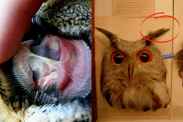
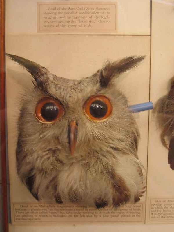
Great Horned Owls and Eagle Owls also show asymmetry, though not as dramatic as the Barn Owl. Their larger skull size helps capture a wider range of sound, which suits their role as powerful, generalist predators. The feather tufts on top of their heads often trick people into thinking they are ears, but they serve no hearing purpose. Instead, these tufts help with camouflage and communication while the real ears remain hidden along the sides of the head.
Snowy Owls live in a completely different world, where the bright, open Arctic landscape means vision plays a larger role in hunting. Because of this, their ears are more symmetrical and less specialized for vertical sound differences. They still have strong hearing, but they rely on it alongside their excellent eyesight rather than as their primary tool.
Great Gray Owls sit at the opposite end of the spectrum. They have some of the largest facial discs of any owl, and this broad sound-collecting surface gives them superb acoustic sensitivity. Their ear placement and facial structure allow them to detect prey moving beneath deep layers of snow, a skill essential for surviving northern winters.
Taken together, these species show how owl ear design shifts in response to the environment. Dense forests and snowy habitats favor highly specialized, asymmetrical ears that can pinpoint sound in three dimensions, while wide, bright landscapes support more balanced hearing paired with sharp eyesight. Each owl’s ear placement reflects exactly what it needs to succeed in its world.

How Owl Hearing Compares to Other Birds
Owls rank among the most skilled listeners in the entire bird world, and their hearing plays a much larger role in their survival than it does for most other species. While many birds depend primarily on vision for hunting, migration, or recognizing mates, owls rely on their ears as their main guide, especially in the dark hours when they do most of their hunting.
One of the biggest differences between owls and other birds is their ability to detect low-frequency sounds. These deeper tones, like the rustle of an animal moving through leaves or the faint shifting of a vole beneath snow, travel well through dense habitats and are easy for an owl to pick up. Most birds are better at hearing higher-pitched notes, which suits communication but does not help much with tracking hidden prey at night.
Owls also outperform other birds when it comes to directional hearing. Their ear openings are often positioned at slightly different heights on the head, which allows them to detect tiny differences in when a sound reaches each ear. This asymmetry gives them an extraordinary sense of direction, letting them pinpoint the exact location of a sound with an accuracy that is closer to what we see in mammals like cats or foxes than in other birds.
In contrast, most bird species have symmetrical ears and lack the pronounced facial disc that funnels sound so effectively into an owl’s ear canals. Without this structure, they cannot create the same three dimensional sound picture that owls rely on for hunting in darkness. Songbirds, for example, excel at recognizing complex high-frequency calls but cannot use sound to map their environment the way an owl can. Even other raptors, such as hawks and eagles, depend far more on vision and have nowhere near the same level of hearing specialization.
These differences explain why owls dominate nighttime hunting niches. They use sound not just as a tool but as their primary sensory system, allowing them to hunt successfully in conditions that leave most other birds at a disadvantage.
Related Post
- Rufous Owl : A to Z Guide
- What Does A White Owl Mean : A to Z Explanation
- Do Owls Eat Birds : Let’s Understand Why
- Baby Great Horned Owl – A to Z Guide
Resources